
Keratoconus Treatment: Cross-Linking, Rings, Lenses or Transplant?
Keratoconus treatment explained by stage: when cross-linking stops progression, when corneal rings or scleral lenses help, and when a corneal transplant is needed. Can keratoconus be cured?
If you have just been told you have keratoconus, the first thing to know is that it can be stopped. What it cannot be, at least not yet, is reversed. Almost every keratoconus treatment falls into one of three jobs: halting the thinning of the cornea, correcting the vision the cone has already distorted, or, in the most advanced cases, replacing the cornea. Which of these you need depends on how far the disease has progressed and how old you are.
We put this guide together because most of the keratoconus patients who contact MPGCARE have received three different opinions and are not sure whether they need cross-linking next month or a transplant next year. Below is how eye surgeons actually decide.
What Is Keratoconus?
Keratoconus is a condition in which the cornea, the clear dome at the front of the eye, gradually thins and bulges outward into a cone shape. A normal cornea is evenly curved, like a ball; a keratoconic cornea is irregular, like the tip of an egg. Light entering the eye is scattered rather than focused, and vision becomes blurred and distorted in a way that glasses cannot fully fix.
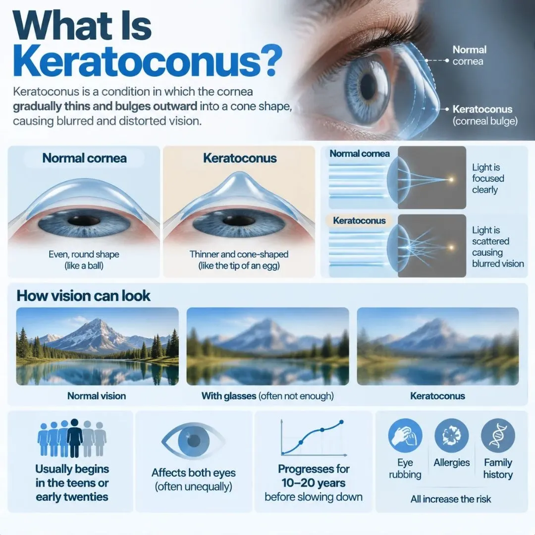
It usually begins in the teens or early twenties, affects both eyes although often unequally, and progresses for ten to twenty years before slowing down. Vigorous eye rubbing, allergies and a family history all raise the risk. Older estimates put keratoconus at one in 2,000 people; modern corneal imaging suggests it is closer to one in 400.
Keratoconus Symptoms and Early Signs
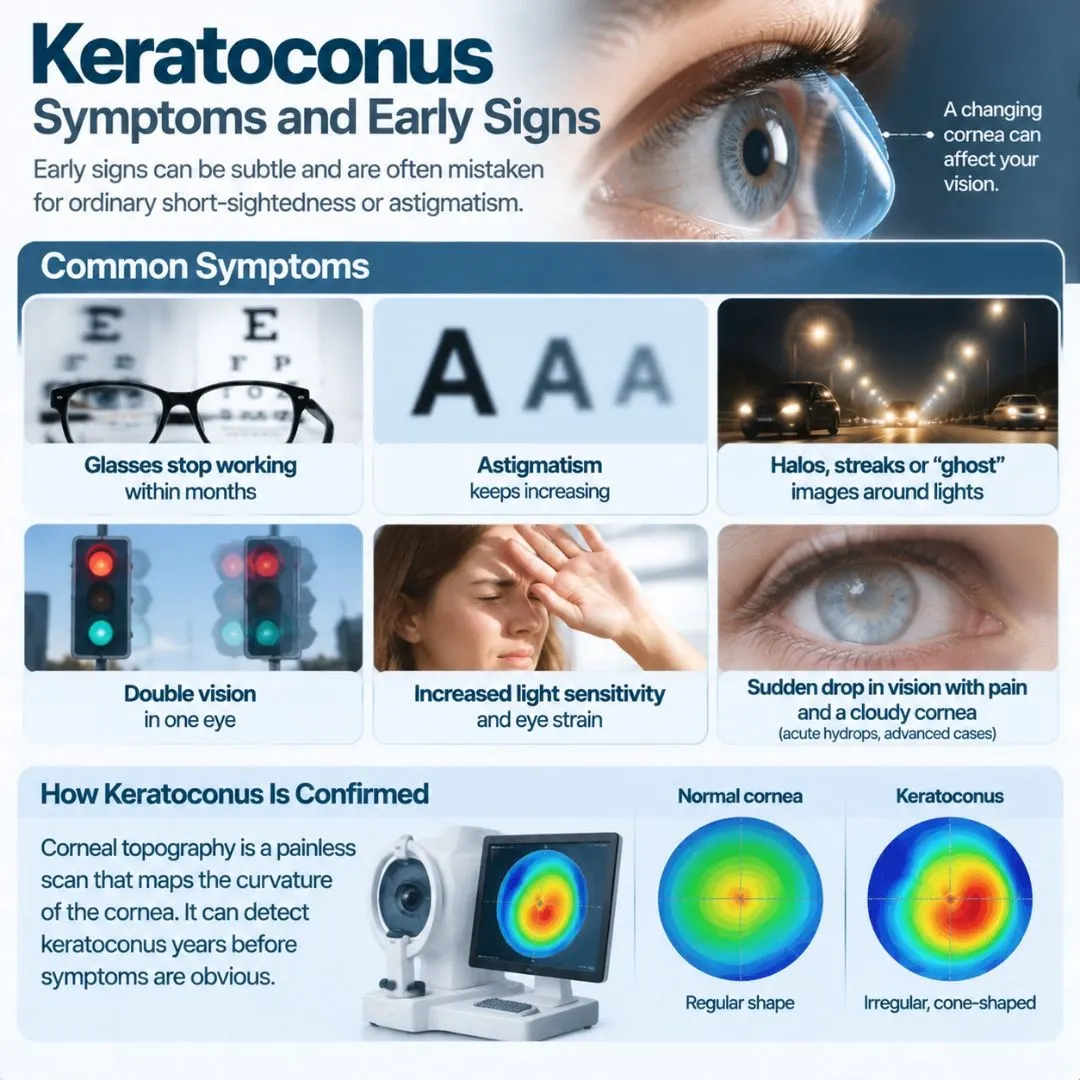
Early keratoconus symptoms are easy to mistake for ordinary short-sightedness or astigmatism. The clues that point to keratoconus rather than a simple prescription change are:
-
glasses that stop working within months of being made
-
astigmatism that increases at every eye test
-
halos, streaks or “ghost” images around lights at night
-
double vision in one eye, even with the other eye closed
-
increasing light sensitivity and eye strain
-
a sudden drop in vision with pain and a cloudy cornea (acute hydrops, in advanced cases)
Because the first sign is often just a rapidly changing prescription, keratoconus is frequently found late. Corneal topography, a painless scan that maps the curvature of the cornea, is the test that confirms it, and it can pick up keratoconus years before symptoms are obvious.
Keratoconus Stages: Mild, Moderate and Advanced
Doctors grade keratoconus using the steepness of the cornea (the K reading, in dioptres), its thickness at the thinnest point (in microns) and how well vision can still be corrected. The classic Amsler-Krumeich system has four stages; in practice most surgeons think in terms of mild, moderate and advanced.
| Keratoconus stage | Corneal steepness (K) | Corneal thickness | Vision | Typical treatment |
|---|---|---|---|---|
| Stage 1 – Mild | Under 48 D | Over 500 µm | Correctable with glasses | Cross-linking if progressing |
| Stage 2 – Moderate | 48–53 D | 400–500 µm | Needs rigid or scleral lenses | Cross-linking + lenses or corneal rings |
| Stage 3 – Advanced | 53–55 D | 300–400 µm | Poor vision even with lenses | Corneal rings, scleral lenses; transplant considered |
| Stage 4 – Severe | Over 55 D, often unreadable | Under 300 µm or corneal scarring | Cannot be corrected | Corneal transplant |
The stage matters because some keratoconus treatments only work within a certain range. Cross-linking, for example, needs a cornea at least about 400 microns thick, which is why acting early keeps more options open.
Can Keratoconus Be Cured?
No. There is currently no keratoconus cure that restores a cornea to its original shape. What treatment can do is stop the disease from getting worse, which in practical terms is almost as good, because keratoconus that has stopped progressing at a mild stage may never need anything more than glasses or lenses.
The distinction is worth holding on to when reading about keratoconus treatment online. Cross-linking strengthens the cornea; it does not thin the cone. Corneal rings and lenses reshape or bypass the cone; they do not remove it. Only a transplant replaces the diseased tissue, and even then the new cornea is a graft, not a cure of the underlying tendency.
Keratoconus Treatment Options by Stage
Every keratoconus treatment does one of three things: it stops progression, it improves vision, or it replaces the cornea. Most patients need a combination over time.
| Goal | Treatment | Best suited to |
|---|---|---|
| Stop progression | Corneal cross-linking (CXL) | Any stage that is still progressing, cornea over ~400 µm |
| Improve vision | Glasses, soft toric lenses | Mild keratoconus |
| Improve vision | Rigid gas-permeable or scleral lenses | Moderate to advanced keratoconus |
| Improve vision | Corneal ring segments (Intacs, Keraring) | Moderate keratoconus, lens-intolerant patients |
| Improve vision | Topography-guided PRK combined with CXL | Mild to moderate, selected cases |
| Replace cornea | DALK or penetrating keratoplasty | Advanced keratoconus, corneal scarring, or lens-intolerant cases |
Corneal Cross-Linking (CXL): Stopping Progression
Corneal cross-linking is the only keratoconus treatment proven to halt progression, and it has changed the outlook for the disease since it was introduced. Riboflavin (vitamin B2) drops are applied to the cornea and then activated with ultraviolet light for several minutes.
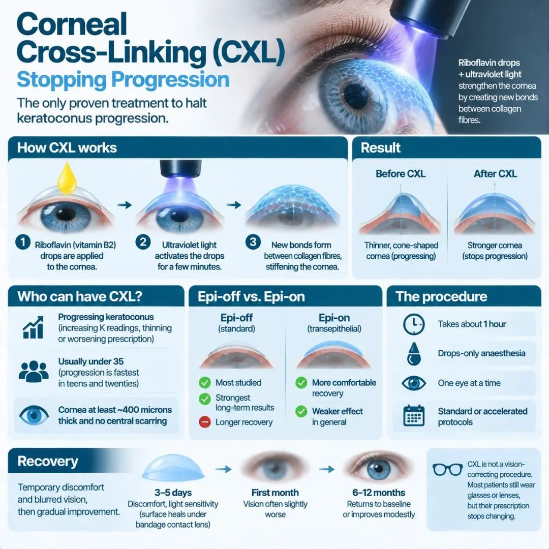
The reaction creates new bonds between the collagen fibres, stiffening the cornea so it stops bulging. The procedure takes about an hour, is done under drops-only anaesthesia, and is performed on one eye at a time.
Who is a candidate for cross-linking?
Anyone whose keratoconus is progressing: a K reading increasing by one dioptre or more within a year, thinning on successive scans, or a worsening prescription. Because progression is fastest in the teens and twenties, most cross-linking is done in patients under 35. The cornea needs to be at least about 400 microns thick at its thinnest point, and there should be no central scarring.
Epi-off vs. epi-on cross-linking
In standard “epi-off” cross-linking, the surface layer of the cornea (the epithelium) is removed so riboflavin can penetrate fully. It is the most studied method and has the strongest long-term results. “Epi-on” (transepithelial) cross-linking leaves the surface intact; recovery is more comfortable, but the effect is generally weaker. Accelerated protocols shorten the ultraviolet exposure without changing the principle.
Recovery after cross-linking
Expect discomfort, light sensitivity and blurred vision for three to five days while the surface heals under a bandage contact lens. Vision is often slightly worse for the first month and then returns to baseline or improves modestly over six to twelve months. Cross-linking is not a vision-correcting procedure; most patients still wear glasses or lenses afterwards, but their prescription stops changing.
Glasses and Contact Lenses for Keratoconus
In mild keratoconus, glasses or soft toric contact lenses may be enough. As the cornea becomes more irregular, they stop working because they cannot correct an uneven surface. That is when specialty lenses take over.
Rigid gas-permeable (RGP) lenses
A rigid lens holds its shape over the cone, creating a smooth new front surface with tears filling the gap underneath. RGP lenses give sharp vision in moderate keratoconus, but they are small, sit on the cornea, and some patients find them uncomfortable.
Scleral lenses
Scleral lenses for keratoconus are larger rigid lenses that vault over the entire cornea and rest on the white of the eye. They do not touch the cone, which makes them far more comfortable, and they correct even advanced irregularity. For many patients with moderate to advanced keratoconus, a well-fitted scleral lens postpones or removes the need for surgery altogether.
Hybrid lenses
Hybrid lenses have a rigid centre with a soft skirt, combining the optics of an RGP with the comfort of a soft lens. They are an option when RGP lenses are intolerable but scleral lenses are not available.
Corneal Ring Segments (Intacs, Keraring)
Corneal ring segments are small, clear crescents of plastic implanted within the cornea to flatten and regularise the cone. Keratoconus ring surgery takes about twenty minutes and is reversible: the segments can be removed or exchanged.
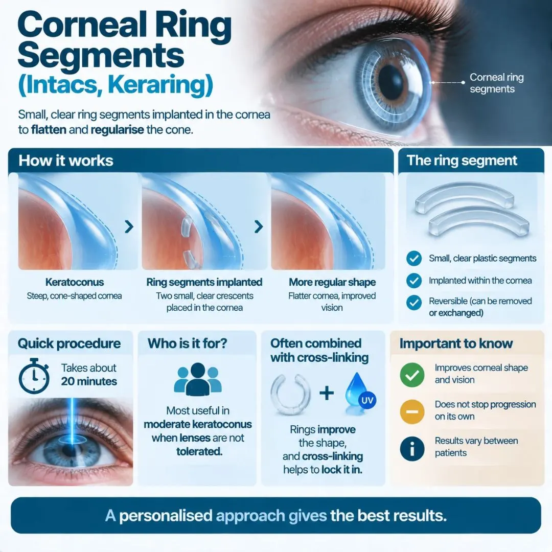
They are most useful in moderate keratoconus when a patient cannot tolerate lenses, and are often combined with cross-linking in the same session, the rings improving the shape and the cross-linking locking it in. Rings do not stop progression on their own, and results vary more than with other treatments.
Corneal Transplant for Keratoconus: DALK vs. PK
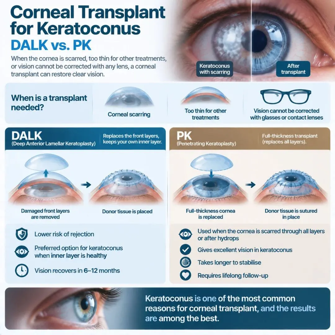
When keratoconus reaches the point where the cornea is scarred, too thin for other treatments, or vision cannot be corrected with any lens, a corneal transplant becomes the answer. Keratoconus is in fact one of the most common reasons for corneal transplant worldwide, and the results are among the best.
DALK (deep anterior lamellar keratoplasty)
DALK replaces the front layers of the cornea while keeping the patient’s own inner endothelium. Because the endothelium is where rejection starts, DALK carries a much lower risk of graft rejection and is the preferred keratoconus surgery whenever the inner layer is healthy. Recovery of vision takes six to twelve months.
Penetrating keratoplasty (PK)
A full-thickness corneal transplant, used when the cornea is scarred through all its layers or after an episode of hydrops. It gives excellent vision in keratoconus but takes longer to stabilise and needs lifelong care. We explain both techniques, recovery and success rates in our guide to corneal transplant in Turkey.
Can You Have LASIK with Keratoconus?
No. LASIK removes corneal tissue and would weaken a cornea that is already thin and unstable; keratoconus, even a suspected early form, is an absolute contraindication. Any laser clinic that offers LASIK without corneal topography is not screening for it properly.
The one exception is a limited, topography-guided surface treatment (PRK) combined with cross-linking in the same session, sometimes called the Athens protocol. It removes only a small amount of tissue to regularise the surface and is suitable for selected mild to moderate cases with enough corneal thickness. It is a keratoconus treatment, not a refractive one, and it is chosen by a corneal specialist, not requested by the patient.
Does Keratoconus Get Worse With Age?
Keratoconus typically progresses from the teens through the twenties and thirties and then slows or stops, because the cornea naturally stiffens with age. After about 40, progression is uncommon, though not impossible, and many patients simply keep the lenses they have.
That is why follow-up is age-dependent. A newly diagnosed teenager is scanned every three to six months; a stable 45-year-old may need a topography once a year or less. If you are under 30, do not wait for symptoms to worsen before asking about cross-linking. Every dioptre of progression that is prevented is vision that does not have to be recovered later.
Keratoconus Treatment in Turkey with MPGCARE
Many of the people who write to us have been told they need cross-linking but are on a waiting list, or have been quoted for scleral lenses they cannot obtain at home. In Turkey, corneal topography, a corneal specialist’s assessment and cross-linking can be completed in a single visit of two to three days, and ring implantation or DALK scheduled within weeks. Scleral lens fitting is available in the same eye centres.
MPGCARE coordinates keratoconus treatment with the ophthalmology departments of accredited partner hospitals, including Liv Hospital Vadi Istanbul, arranges flights, accommodation and transfers with an interpreter, and can add a general health check-up if you want to use the trip fully.
Send your most recent topography or prescription through our online assessment form and a corneal specialist will tell you, before you book anything, whether cross-linking, lenses, rings or a transplant is the right next step. If you are already at the transplant stage, our corneal transplant guide covers what to expect.
Frequently Asked Questions
Is keratoconus a serious eye condition?
It is serious in the sense that untreated progressive keratoconus can lead to severe vision loss, but with cross-linking and modern lenses most patients keep good, functional vision for life. The key is catching it while it is still progressing.
What happens if keratoconus is left untreated?
The cornea continues to thin and steepen, glasses and then lenses stop working, and scarring or hydrops can develop. A significant proportion of untreated advanced cases eventually need a corneal transplant, which cross-linking at an earlier stage would have prevented in most of them.
How long does cross-linking last?
Long-term studies show the effect holding for ten years and beyond in the great majority of patients. A small number, mostly the very young, progress again and can be re-treated.
Can keratoconus cause blindness?
Total blindness is very rare. Keratoconus affects only the cornea, and even in the most advanced cases a corneal transplant restores vision. It can, however, cause severe visual impairment if it progresses untreated.
Is keratoconus hereditary?
Partly. Around one in ten patients has a close relative with keratoconus, and it is more common in some families and populations. If you have it, your children should have corneal topography in their early teens.
What is the best treatment for keratoconus?
There is no single best keratoconus treatment. For a progressing cornea it is cross-linking; for correcting vision it is usually scleral lenses; for an advanced, scarred cornea it is DALK. Most patients need two of these over their lifetime, in that order.
This article is for general information and does not replace an examination by a corneal specialist. Treatment choice depends on your corneal topography, thickness and rate of progression.